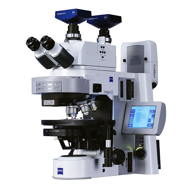
example picture
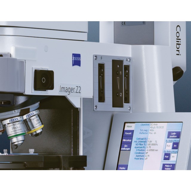

Axio Imager.Z2
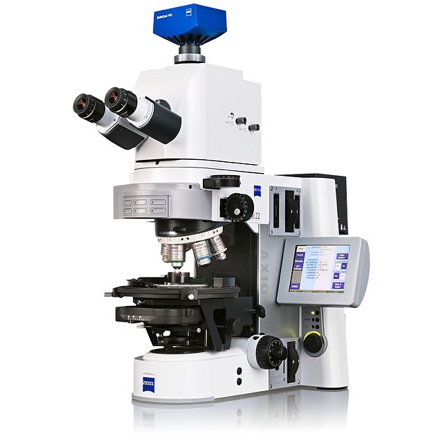
example picture
Axio Imager.Z2
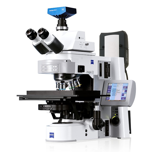
example picture
Axio Imager.Z2
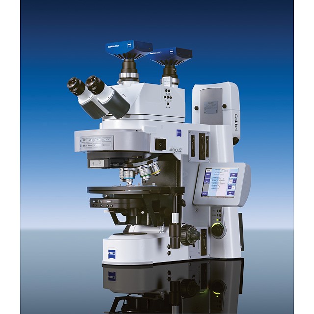
example picture
Axio Imager.Z2
example picture
Axio Imager.Z2
example picture
ZEISS Microscopy How-to: Set up Köhler Illumination on your ZEISS Axio Imager.Z2
Axio Imager scan Advanced
Item no.: 000000-2647-163 (individual configuration)
Description
Full description
Promotion WF Axio Imager.Z2 for Screening - Advanced 8-slide stage, Colibri 7 (7 LEDs) , Apotome 3, ...
Axio Imager scan Advanced
Item Number: 000000-2647-163 (individual configuration) Reset configuration
Please note that if an item is removed from or added to a preconfigured kit the full functionality can not be guaranteed!
The current configuration contains no more items and can not be added to the shopping cart.
Current configuration
-
Axio Imager.Z2 microscope stand with Z-drive mot. and TFT monitor
Item no.: 430000-9902-000 -
Fine drive disk, flat with scala for Axio Imager 2, changeable
Item no.: 430051-9010-000 -
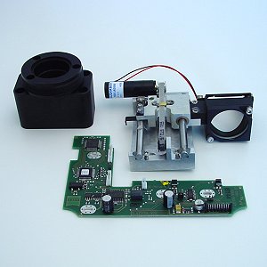
Camera path deflection on the left side, mot., interface 60N
Item no.: 425104-0000-000 -
Path deflecting mirror 100% for camera deflection, 34x46x4 mm
Item no.: 425110-9110-000
-
6-position objective nosepiece, HD DIC M27 mot.
Item no.: 424505-0000-000 -
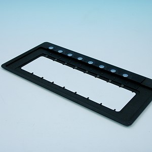
Transmitted-light/reflected-light stage carrier, detachable
Item no.: 430704-9901-000 -
Condenser carrier with vertical adjustment on both sides
Item no.: 430705-0000-000 -
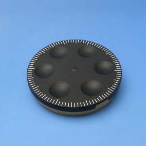
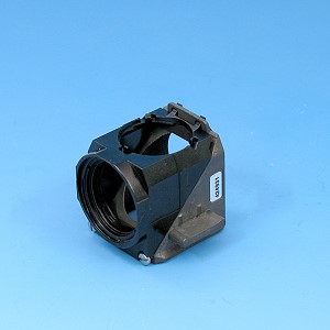
10-position reflector turret mot. ACR, for P&C modules
Item no.: 424913-0000-000 -
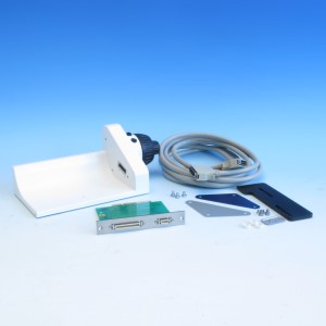
Docking station for TFT-Display
Item no.: 432907-9901-000
-
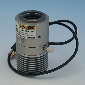
Attachment lamp VIS-LED with collector
Item no.: 423053-9030-000
-
Solid-State Light Source Colibri 7, Type FR-R[G/Y]BV-UV
Item no.: 423052-9770-000
-
Binocular phototube 15°/25 (100:0/0:100), upright image
Item no.: 425503-9901-000
-
Filter set 70 HE Alexa 430 shift free (E)
Item no.: 489070-0000-000 -
Filter Set 96 HE BFP shift free (E)
Item no.: 489096-9100-000 -
Filter set 43 HE Cy 3 shift free (E)
Item no.: 489043-9901-000 -
Filter set 38 HE eGFP shift free (E)
Item no.: 489038-9901-000 -
Filter set 50 Cy 5 shift free (E)
Item no.: 488050-9901-000 -
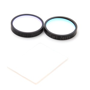
Filter set 64 HE mPlum shift free (E)
Item no.: 489064-0000-000 -
Filter Set 112 HE LED (E)
Item no.: 489112-9110-000
-
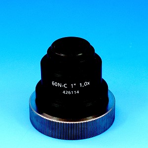
Camera Adapter 60N-C 1" 1.0x
Item no.: 426114-0000-000
-
Eyepiece eyecup
Item no.: 444801-0000-000
-
Universal condenser achromatic-aplanatic 0.9 H D Ph DIC, mot.
Item no.: 424201-9903-000
-
Graphics Card NVIDIA RTX A4000 16 GB DP (O)
Item no.: 410331-2800-000 -
Monitor TFT 27" HP Z27q G3 (68 cm) (O)
Item no.: 410350-2702-000 -
Language Pack English US (O)
Item no.: 410380-0200-000 -
Network Adapter 2 x 10 GbE RJ45 (HP Z8 G5) (O)
Item no.: 410358-0505-000 -
Memory 32 GB (1x32) DDR5 4800 MHz ECC reg RAM (HP Z8 G5) (O)
Item no.: 410303-3206-000 -
Processor Intel Xeon W7-3455 (HP Z8 G5) (O)
Item no.: 410330-1200-000 -
System Configuration 2 for HP Z8 G5 (O)
Item no.: 000000-2615-590 -
Microscopy Workstation Premium (HP Z8 G5) (O)
Item no.: 410203-9914-000 -
30m Patch Cable CAT7
Item no.: 000000-2147-606 -
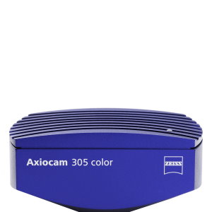
Digital Microscopy Camera Axiocam 305 color R2 (D)
Item no.: 426560-9031-000 -
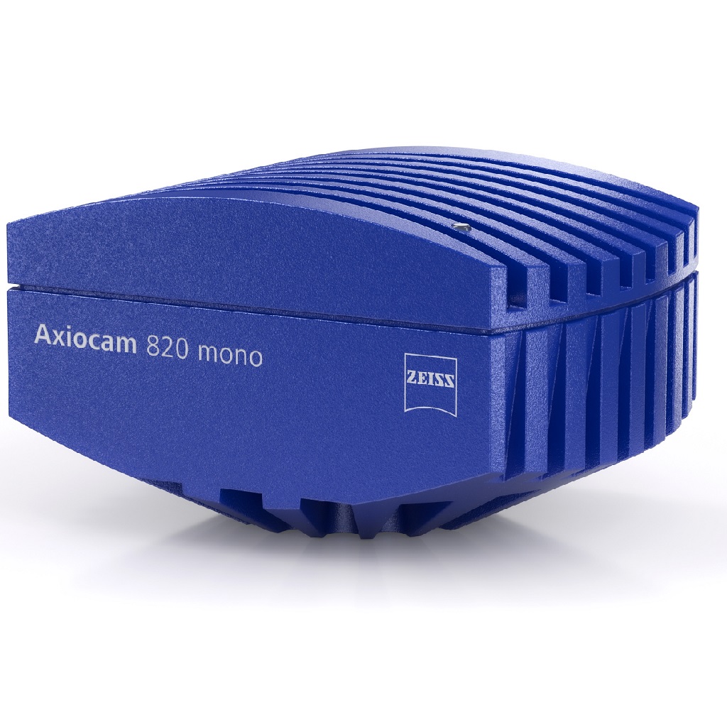
Microscopy Camera Axiocam 820 mono
Item no.: 426560-9190-000
-
ZEN 3.9
Item no.: 410135-0002-390 -
ZEN Toolkit Motorized Acquisition
Item no.: 410136-0165-390 -
ZEN Toolkit Smart Acquisition
Item no.: 410136-0167-360 -
ZEN Toolkit 2D
Item no.: 410136-0168-360 -
ZEN Toolkit Bio Apps
Item no.: 410136-0175-370 -
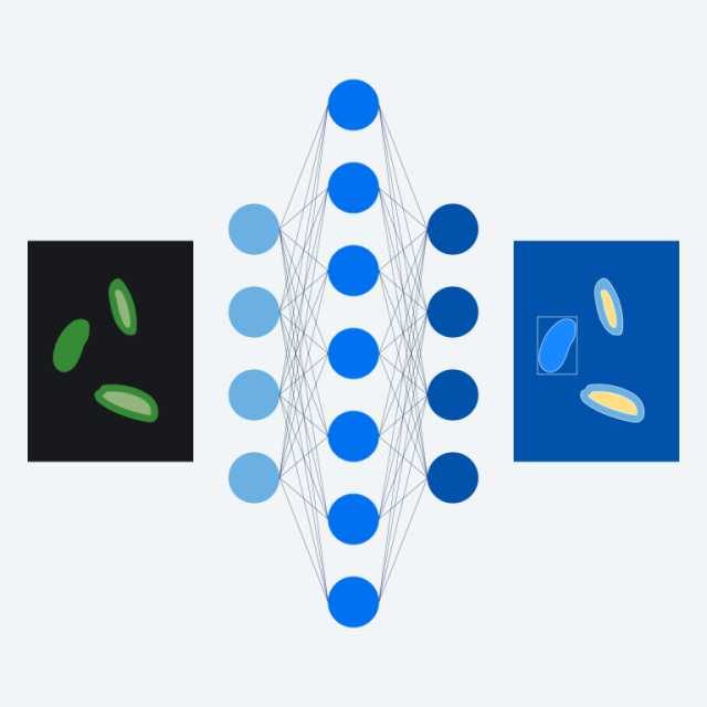
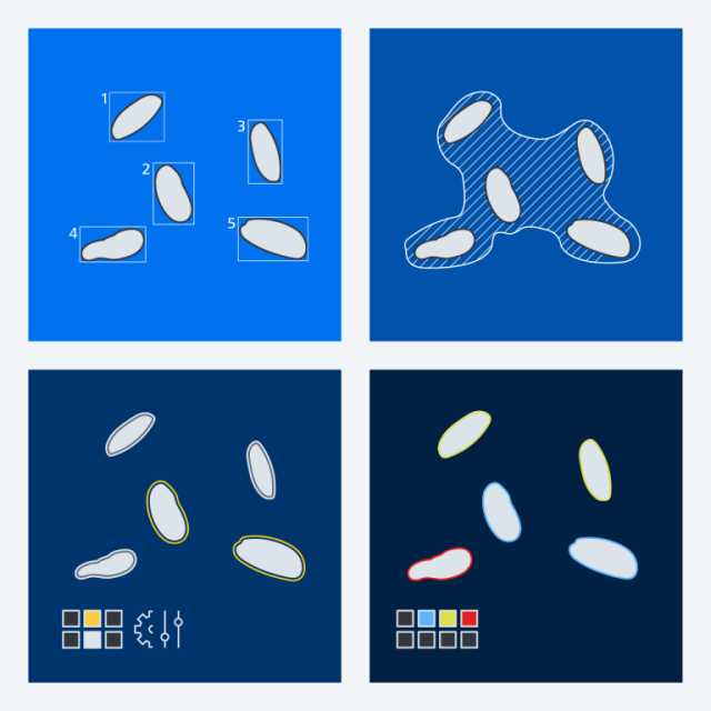
ZEN Toolkit AI
Item no.: 410136-0169-380
-
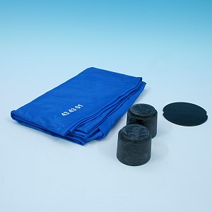
Dust protection Set L
Item no.: 434304-0000-000
-
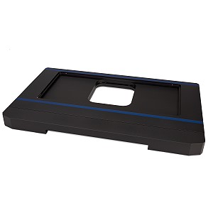
Anti vibration plate Axio Imager and LSM 800/900(D)
Item no.: 400102-8311-000
There is no parts list for this product.